Advertisement

Archives

gravatar

Acute Pain Service(APS)


Contents
1)Methods of analgesia
2)Common drugs
3)Specific drug

METHODS OF ANALGESIA
1)PRE-EMPTIVE ANALGESIA
2)COMBINATION ANALGESIA
3)INTERRUPTION AT SPECIFIC SITES OF PAIN TRANSMISSION

PRE-EMPTIVE ANALGESIA
1) Analgesic drugs are given before any surgical incision
2) Concept that suppression of dorsal horn neuronal activity involved in pain pathway before painful stimulus (e.g. surgery)
3) It may reduce- operative pain -as evidenced by delayed request for additional post-op analgesia-doses of analgesia-pain scores
4) Techniques of suppression – local anaesthetic , epidural , spinal anaesthesia, NSAID and opiod analgesic.

COMBINATION
1)Opioids
2)NSAIDs
3)Nerve blocks

INTERRUPTION AT SPECIFIC SITES OF PAIN TRANSMISSION
1) REDUCED PERIPHERAL STIMULUS
e.g. NSAIDs
2)PERIPHERAL PAIN
.g. LA
3)CENTRAL PAIN TRANSMISSION
e.g.EPIDURAL
4)STIMULATION OF INHIBITORY PATHWAYS
e.g. opioids, acupuncture

COMMON DRUGS
1)OPIOIDS
morphine
pethidine
fentanyl
nubain

2)LOCAL ANAESTHETICS
lignocaine (lidocaine)
marcaine(bupivacaine)
Naropin (ropivacaine)

3)OTHERS
NSAIDs
Paracetamol

COMMON TECHNIQUES
1) SUBCUTANEOUS
- Morphine
2) PCA
-morphine
-pethidine
3) EPIDURAL ANALGESIA
-pethidine
- “LA + opioid cocktail”
-PCEA with pethidine
4) IV INFUSION
-morphine
-pethidine
-nubain
5) ITM(Intrathecal Morphine)
6) Oral/Supp. PCM/NSAIDs
(oral, supp, IM or IV)

SPECIFIC DRUGS
OPIOIDS
Narcotic - Greek narco, meaning deaden or numb
Morphine - Greek Morpheus, god of dreams, son of the god of sleep
Opium - is obtained from the unripe seed capsule of the poppy plant
Opiates - drugs derived from opium
Opioids - refers to all drugs natural & synthetic, that has morphine like properties and act through the opioid receptors in the body

NATURALLY OCCURRING:
1)MORPHINE
2)CODEINE
3)PAPAVERATUM (OMNOPON)

SYNTHETIC:
1)DIAMORPHINE (HEROIN)2)PETHIDINE3)FENTANYL4)NALBUPHINE(NUBAIN)5)TRAMADOL (TRAMAL)

SIDE EFFECTS
1) Sedation, euphoria,miosis
2)Respiratory depression
3)Nausea & Vomiting
4)Gastric stasis, ileus
5)Constipation
6)Urinary retentionPruritus

OPIOIDS : Morphine
Standard opioid
Clinically superior in relieving pain
Uses:moderate to severe pain, cancer
R&D: IM,SC: 0.1-0.2mg/kg & IV : 0.05-0.2mg/kg 4 hourly inf: 10-40mcg/kg/hr (adult/paed)ITM: 0.2mg
Peak effect: 20min(IV), 30-60min(IM)

OPIOIDS : Pethidine
-10 X less potent than morphine (100mg pethidine = 10 mg morphine)
-For moderate to severe pain
-R&D: Parenteral- 0.5-1.0mg/kg
infusion: 100-300mcg/kg/hr
epidural: 25mg or infusion: 10mg/hr
-Peak effect: 10 min(IV), 20-30 min(IM)

OPIOIDS : Fentanyl
-100 X more potent than morphine(0.1mg fentanyl = 10 mg morphine)
-Dose: 1 – 2 mcg/kg
-Duration: 30 – 60 min
-Uses: mainly for intraoperative analgesia as well as in epidural cocktails

OPIOIDS : Nubain
-Agonist-antagonist property
-Uses: moderate to severe pain
-R&D: IV,IM,SC :10-20mg 3-6 hourly
children :0.2-0.5mg/kg/dose
-Peak effect: 30min (IM)
-Duration: 3 – 6 hours

LOCAL ANAESTHETIC

2 type of Local Anaesthetic

1)AMIDE
LIGNOCAINE (XYLOCAINE)
BUPIVACAINE (MARCAIN)
ROPIVACAINE (NAROPIN)

2)ESTER
COCAINE

MODE OF ACTION
Block nerve conduction, prevent action potential by binding to Na+ channels

LA:Effects on organs
1)CVS : arrhythmias, hypotension, bradycardia, cardiac arrest
2)Resp.: depresses hypoxic drive, apnea
3)CNS: early:- circumoral numbness, tongue paraesthesia, dizziness,tinnitus, blurred vision
4)Excitatory signs: restless, agitation, nervousness
5)CNS depression: drowsiness, coma

THE RELATIONSHIP BETWEEN LIGNOCAINE PLASMA CONCENTRATION AND PHARMACOLOGICAL EFFECTS

PLASMA CONCENTRATION(mg/ml)

26 CVS depression
24
22
20 Resp arrest
16 coma
12 unconciousness
10 convulsions
8 muscular twitching
6 visual disturbances
4 lightheadness,tinnitus circumoralnumbness
2 antiarrhymias

LIGNOCAINE (common LA)
PREPARATION
: LIGNOCAINE HCL0.5/1.0/1.5/2.0%
MAXIMUM DOSE : 5 mg/kg (plain)7 mg/kg (+ adr.)

BUPIVACAINE
PREPARATION: BUPIVACAINE HCL0.25 / 0.5 / 0.75%

EMLA 5%
-Topical local anaesthetic
-Eutectic Mixture of Local Anaesthetics
-An oil/water emulsion of lignocaine & prilocaine (1:1), melting point at 18ºC, liquid at room temp., penetrate the skin.
-Optimum effect : after a minimum of 60 min of application.1.5-2.0gm/10m2
-Duration: 30min.
-S/E: transient paleness or redness of skin, itchy, methaemoglobinemia-CAUTION:infant less than a year

NSAIDs

Mode of Action: by inhibiting the enzyme cyclooxygenase which is involved in the synthesis of PGs in the body (PGs- play a part in the transmission of pain)
Analgesic, anti-inflammatory & anti- pyretic action

Route: Oral : Mefenamic acid(ponstan), Naproxen sodium (synflex), Indomethacin, Diclofenac (voltaren), Vioxx, Celebrex. IM: Diclofenac, ketorolac & orudis IM&IV

SIDE EFFECTS
1) GIT irritation2) antiplatelet activity3) decrease renal blood flow, may precipitate ARF4) Allergic reactions

CONTRAINDICATIONS
1) H/O coagulopathy
2) peptic ulcer
3) renal impairment
4) H/O allergy to aspirin or other NSAIDs

PARACETAMOL(PANADOL)
An acetanilide derivative
Uses: an analgesic for mild to moderate pain & antipyretic
Preparation: tablet 500mg, supp.:125/250/500mg, syrup: 125/250/500mg per ml
Dosage: Adult (O&R) 500-1000mg 4-6hourly. Child (O) 10-15mg/kg (R) 15-20mg/kg 4-6hrly
S/E: GIT disturbances, hepatic necrosis (15gram/dose), renal failure, hypoglycemia

gravatar

MANAGING MYOCARDIAL INFARCTION-The role of ECG


P - atrial electrical activation
PR-Interval bet. atrial and ventricular activation
QRS - ventricular electrical activation
ST-interval bet.end of ventricular activation and ventricular recovery
T- ventricular recovery
U - ? Purkinje fibers recovery
QT- total duration of ventricular activation & recovery


P : upright in I,II, V2-V6 , ht <>
Q : Ht <>
QRS : <>
ST segment : isoelectric or <>
T: upright I,II, V3*, V4-V6
QTc =QT / ÖRR ( €<0.39s>

ST deviation : measure 0.08s ( 2 small boxes ) from J point

ECG IN ST ELEVATION MI

COMMON MISTAKES
- Poor interpretation of ECG changes in STEMI
- Failure to identify concomitant ECG changes / complications
e.g. – arrhythmias, conduction disturbances,RV and posterior MI changes
- Failure to do right sided ECG in inferior MI
- Failure to repeat / monitor ECG when earlier ECGs were normal
- Delay in performing / review ECGs
- Failure to compare earlier/old ECGs if available
- Failure to write name, date and time of ECG performed
- Never assess objectively ST segment elevation pre and post reperfusion therapy


ROLES OF 12 LEAD ECG IN STEMI
- Diagnosis of MI
- Correlate ECG changes and candidate for reperfusion therapy
- Identify location , extent of MI including RV and posterior MI .
- Identify conduction and rhythm disturbances
- Assess the success of reperfusion therapy

Diagnosis
The diagnosis of MI is based on the presence of at least two of the following three criteria:
(1) a clinical history of ischemic-type chest discomfort
(2) changes on serially obtained electrocardiographic tracings
(3) a rise and fall in serum cardiac markers

ECG DIAGNOSIS
A. ST elevation in 2 or more contiguous leads of standard 12 lead ECG
- ≥ 1mm limb leads ( II , III , AVF, AVL , I )
- ≥ 2mm precordial leads ( V1 – V6 )
- WITH OR WITHOUT Q WAVE
B. New onset of LBBB
C. ECG changes of RV INFARCTION – IN RIGHT SIDED
ECG OF V4 /V5
D. ECG changes of POSTERIOR INFARCTION – V1 AND V2

ECG : Evolutionary changes
Hyperacute T waves
•Present for only five to thirty minutes after onset of MI
•T waves more prominent, symmetrical and pointed.

ST segment changes
–Within hours of symptoms
–ST segment straighten, with loss of ST-T wave angle. Then T wave becomes broad and ST segment elevates, losing its normal concavity. As further elevation occurs, the ST segment tends to become convex upward.

Pathological Q waves- Evidence of myocardial necrosis
•Usually develop after 9 hours, occasionally after 24 hours.
-Loss of R wave after 12 hours


Ischemia

•T wave inversion, ST segment depression
•Acute injury: ST segment elevation
•Dead tissue: Q wave
ECG : Evolutionary changes

Resolution of changes in ST segment and T waves.
ST segment elevation diminishes and T wave inverted.
Persistent ST elevation- Possible LV aneurysm

TEMPORAL RELATIONSHIP


gravatar

INTUBATION


ANESTHESIS SHOULD KNOW
1) Anatomy, nerve innervation,phatologic cond. & method of assessment.
2) Physiologic consequences & complication.
3) Anticipate pt. with difficult airway.
4) Formulate & implement alternative plan in various clinical situation.
History
Sn/Sm suggest abnormal airway
•SOB
•Hoarsenes ( rheumatoid artritis- criocotenoid cartilage – narrowing Lx opening.)
Previous surgery, trauma, neoplasia of airway
Previous anesthetic record.

Physical examination
1) Head shape- micrognathia,mandibular hypoplasia
2) protruding teeth(edentulous)- disturb seal mask, loose, capped, prosthetic teeth.
3) Cleft, high-arch palate
4) Large big tongue( trisomy 21, mucopolysaccharidoses, beckwith-weidemann syndrom)
5) Prediction difficulty trachea intubation by:
•Mallampati classification
•Temporomandibular joint mobility
( ankylosis,long term type1 DM, pain due to trauma)
•Distance lower border mandible to thyroid notch > 6.5 cm (adult)

INDICATIONS OF INTUBATION
1) Inabiliy to oxygenate patient (SpO2 <>
2) Inability to ventilate patient (rising PaCO2, respiratory acidosis, mental status change or other symptoms)
3) Patient unable to protect the airway.

CONTRAINDICATIONS
1) Neck immobility or increased risk of neck trauma (e.g. rheumatoid arthritis, cervical spine injury, etc.)-consider fiberoptic intubation.
2) Inability to open mouth (e.g. trismus, scleroderma, surgical wiring, etc.)- consider nasal intubation, either blind or fiberoptic, or surgical airway.

Airway equipment
1) Mask
2) Airways
3) Laryngoscopes
4) Endotracheal tubes + stylet.
•Adult male: 7.0,7.5,8.0
•Adult female: 6.5,7.0,7.5
•Children > 1 year age
size= (age(yr) +16 )/ 4
5) Length of tube cm= 12 + (age/2)
6) Suction.


endotracheal tube


laryngoscope


type of larynscope

Technique For Orotracheal Intubation
- Assemble equipment
- Calculate doses and draw medications into syringes
- Check IV access and flush fluid
- Preparation And Equipment (Always Have Suction Available)
- Head Position - Alignment Of Oral, Pharyngeal, and Laryngeal Axes ,(sniffing position)
- Choice Of Laryngoscope And Endotracheal Tube

Induction agent

Etomidate
dose- 0.3 mg/kg
advantage-Good for low blood pressure; okay in hypovolemia
cautions- Nausea and vomiting on emergence


Ketamine
dose- 1.5 mg/kg
advantage- Good for low blood pressure, hypovolemia; good in asthma
Caution in elevated intracranial pressure or heart disease


Propofol
dose- 2 - 2.5 mg/kg
advantage- Rapid onset and recovery
Caution if hypovolemic or risk of hypotension


Thiopental
dose- 3 - 5 mg/kg
advantage- Multiple drug interactions
caution if hypovolemic or risk of hypotension

Muscle relaxant

Succinylcholine
dose 1 - 1.5 mg/kg
characteristic- Rapid onset, rapid recovery; fasciculation
Contraindicated in hyperkalemia, crush injury, renal failure, extensive burns, elevated intracranial or intraocular pressure

Rocuronium
dose 0.6- 1.2 mg/kg
characteristic- No fasciculation
cautions- Longer acting-may be problematic if intubation attempt fails

Vecuronium
dose 0.08 - 0.1 mg/kg

Atracurium
dose 0.4 - 0.5 mg/kg

Orotracheal Intubation
- Patient’s Head At The Level Of The Xiphoid
- Sniffing Position
- Laryngoscope In LEFT Hand
- Open Mouth
- Hold Tracheal Tube In Right Hand Like A Pencil

Laryngoscopic View Grades
Laryngoscopic view may be clasified into four grades:
Grade I = visualization of the entire laryngeal aperture.


Grade II = visualization of just the posterior portion of the laryngeal aperture.

Grade III = visualization of only the epiglottis.
Grade IV = visualization of just the soft palate only, not even the epiglottis is visible.

Laryngoscopic View Grades
Grade II or III laryngoscopic views are relatively common and occur in 1 to 18% of surgical patients. The Grade III view occurs in about 1-4% of patients. A severe grade III or grade IV view with failed endotracheal intubation occurs 0.05 of 0.35% of patients.
It may be helpful to the next anesthesiologist to record both the grade of laryngoscopic view achieved, the patient position and the technique.


Laryngoscopic View Grades

Alternatives To Orotracheal Intubation Under Anesthesia
Awake Orotracheal Intubation
Nasotracheal Intubation - Awake Blind Nasal - Nasotracheal Intubation After Induction
Intubation With Fiberoptic Bronchoscope
- Awake versus Under Anesthesia
- Orotracheal versus Nasotracheal
Retrograde Intubation

Verification Of Correct Tube Placement
- The most reliable method for determination of tube placement is direct vision of the endotracheal tube passed through the vocal cords
- Symmetric Chest Movement
- Symmetric Breath Sounds
- End tidal Carbon Dioxide - Greater Than 30 For 3-5 Breaths
- Condensation Of Water In The tube
- Palpation Of Cuff In Suprasternal Notch
- Fiberoptic Bronchoscopy


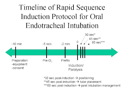
RAPID SEQUENCE INDUCTION
Sequence and rapid method of securing airway, to minimizes aspiration in process of intubation
•Pt preoxygenated for 5 minutes / 4 max deep breath within 30 scd/ 3 vital capacity breath 100% O2.
•No assisted breathing until airway isolated
•Nondepolarizing MR - scolin
•Applied cricoid pressure (sellick’s maneuver)
*All process should be completed in 60 sc post induction

Complication of endotracheal intubation
During intubation
•Laryngospasm
•Laceration,bruising lips,tongue,pharynx
•#,chipping,dislogement of teeth
•Perforation trachea,esophagus
•Retropharyngeal dissection
•# or dislocation cervical spine
•Trauma to eyes
•Haemorrage
•Bactremia
•Aspiration gastric content/ FB
-Endobrachial or esophageal intubation
-Dislocation arytenoid cartilages or mandible
-Hypoxemia, hypercarbia.
-Bradycardia, tachycardia
-Hypertension
-Increased intracranial or intraocular pressure

Complication with tube insitu
-Accidental extubation
-Endobrachial intubation
-Obstruction / kinking
-Bronchospasm
-Ignation of tube by laser device
-Aspiration
-Sinusitis
-Excoriation of nose or mouth

Complication after extubation
-Laryngospasm
-Aspiration –gastric content, blood, FB
-Glottic,subglottic, uvular edema
-Dysphonia,aphonia
-Paralysis of vocal cord, hypoglossal,lingual nerves
-Sore throat
-Noncardiogenic pulmonary edema
-Laryngeal incompetance.
-Soreness, dislocation jaw
-Tracheomalacia
-Glottic,subglottic or tracheal stenosis
-Vocal cord granulomata.


Members of